
Home / Research

Research programme

Four questions we keep returning to.

Each of our lines of work sits at the interface between tumour biology and the cerebral vasculature — the tissue that decides whether a therapy ever reaches its target.

01 — Brain metastasis

The brain changes before the tumour arrives.

A metastasis does not simply land in healthy tissue. The brain is altered in advance — the pre-metastatic niche — and understanding that preparation is the best chance we have of interrupting it.

We use patient-derived tissue from our own HiRes study together with mouse models to analyse metastatic and pre-metastatic tissue at single-cell resolution, comparing human and murine data directly. The goal is a set of biomarkers that predict — and eventually help prevent — brain and bone metastasis.

Alongside this, we ask how the clinical route a sample travels affects what we can learn from it: which preoperative patient parameters shape the quality of surgically obtained brain-metastasis tissue for translational research.

Single-cellPatient tissue Pre-metastatic nicheBiomarkers
Related publications
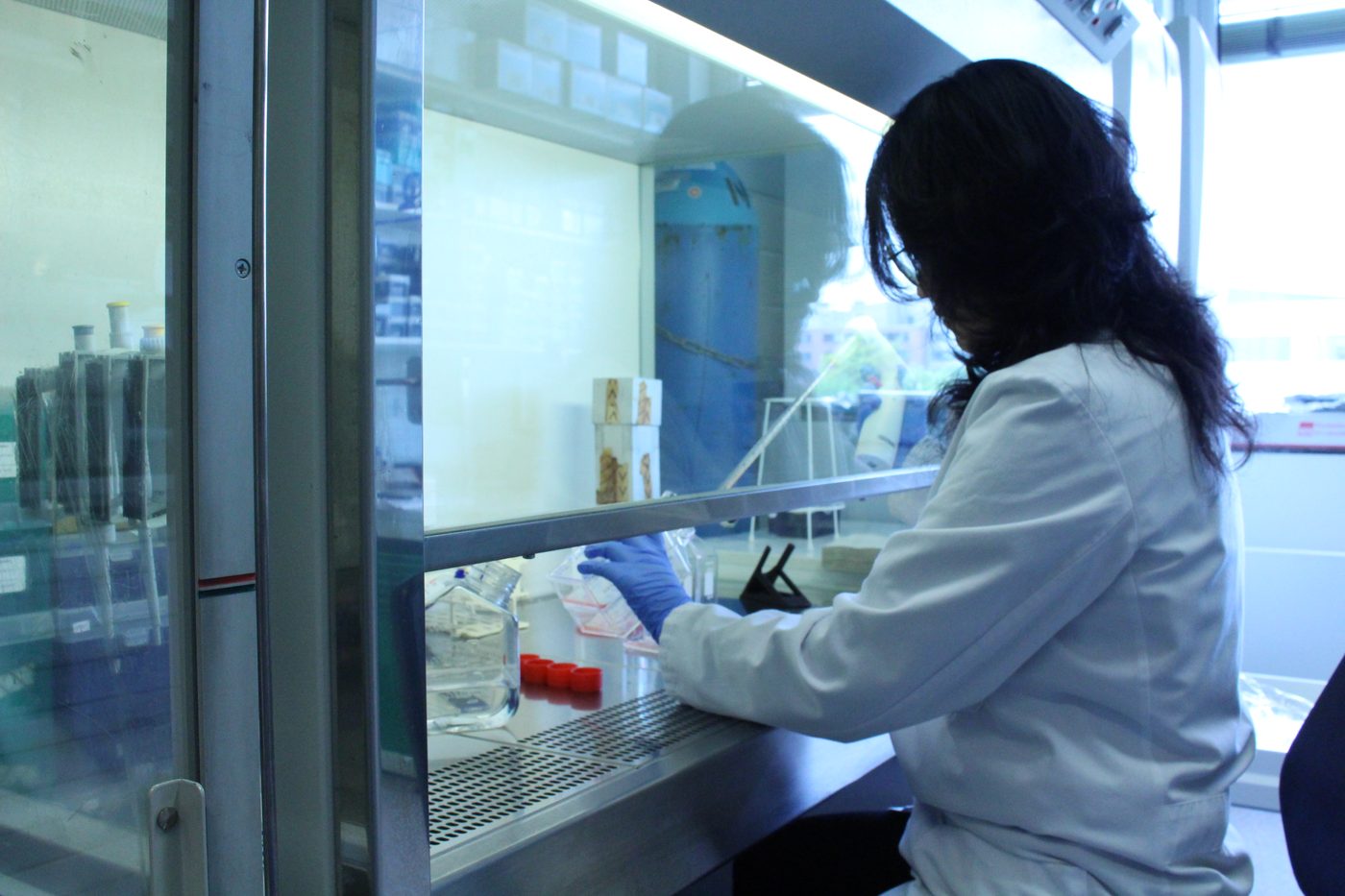
Researcher handling patient-derived tissue samples in a laminar flow hood
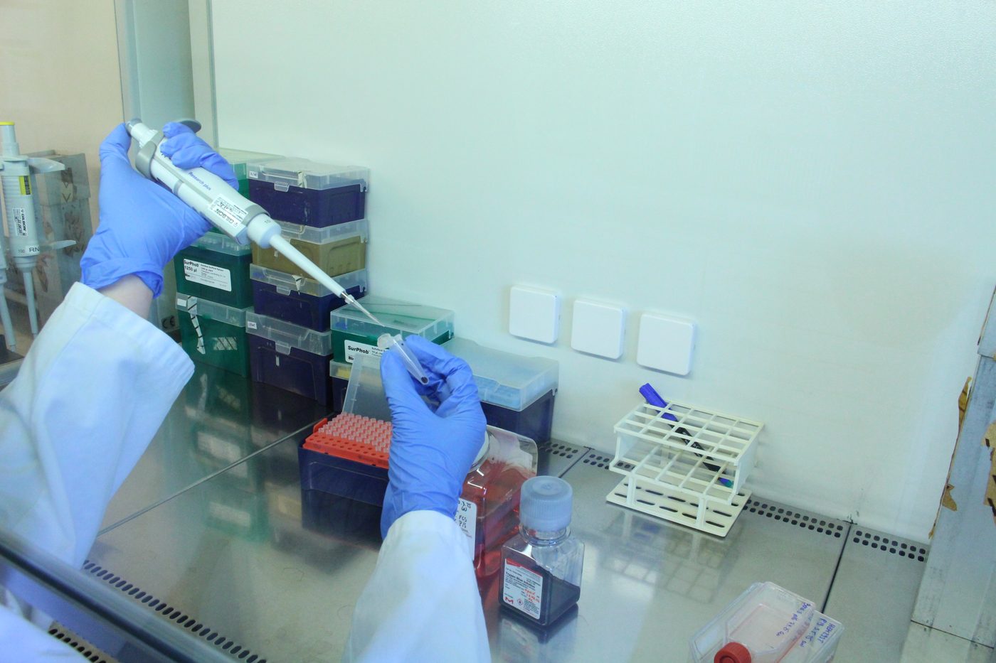
Pipetting under a laminar flow hood during a cell-culture experiment

02 — Glioma

Anti-angiogenic therapy works, until it does not.

Gliomas find their way around treatments that target their blood supply. We want to know which vessels have to change for a drug to keep working — and which changes let the tumour escape.

Our long-running work centres on Ephrin-B2 / EphB4 signalling between tumour and endothelium: how it shapes vascular architecture, how endothelial EphrinB2 governs the response to sunitinib, and how EphB4 mediates resistance to anti-angiogenic therapy in experimental glioma.

We have also examined how tumour vascularisation and the microenvironment — NDRG1 expression, CCR2-positive cells, plasticity-related genes — determine whether a glioma responds to treatment at all.

EphrinB2 · EphB4Anti-angiogenics GlioblastomaTumour microenvironment
Related publications

03 — Neurovascular interface

Waves that travel through the cortex every ten seconds.

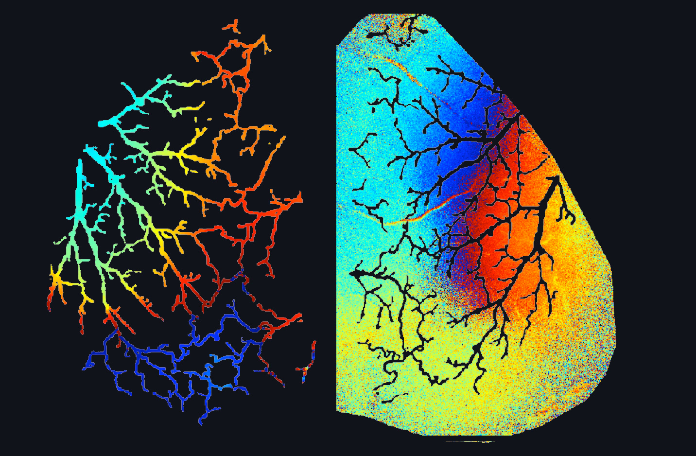
Arterioles oscillate independently of the heartbeat and of brain activity. We showed these oscillations form long-wavelength traveling waves that modulate how the cortex is perfused.

Measured in the penetrating arterioles of the mouse cortex, vasomotion alters blood flow by around 20% — and the effect is stronger at rest than in active brain regions. The waves run along every arteriole at roughly two millimetres per second, in many directions at once. The bulges they raise on the vessel wall may help mix the cerebrospinal fluid, and so improve the clearance of misfolded proteins and metabolic waste.

We are now asking how these waves are altered in stroke, cerebral haemorrhage and neurodegenerative disease — and whether modulating vasomotion could become a therapy in its own right.

Cerebral blood flowTraveling waves fMRI biologyWaste clearance
Read the Neuron paper
Phase map of cortical vasomotion showing traveling waves across the arteriolar network
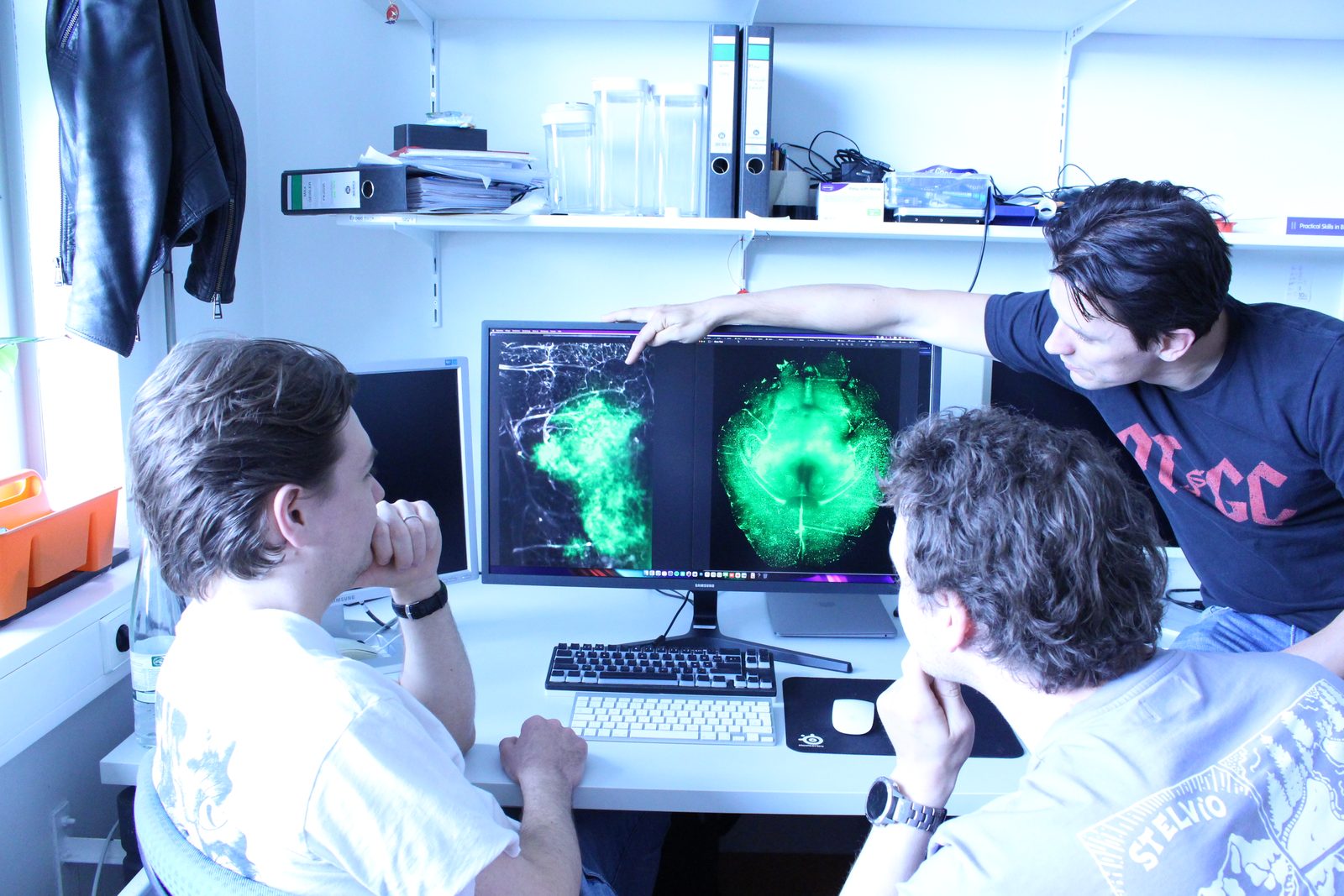
Researchers reviewing fluorescence imaging data of cerebral vasculature

04 — Methods

Better answers need better instruments.

A good deal of our work is building the tools that make the other three questions answerable at all.

We construct and apply adaptive-optics two-photon microscopes with direct wavefront sensing, prepare chronic and lateral cranial windows for long-term observation in the living cortex, and combine wide-field and confocal imaging with small-animal microsurgery to watch blood flow and tumour growth as they happen.

Method development runs the other way too: we contribute to analysis tools — including morphology-free functional fluorescence imaging analysis — so the data these microscopes produce can actually be interpreted.

Two-photonAdaptive optics Cranial windowsImage analysis
Related publications

Infrastructure

What we work with.

The laboratory runs its own imaging, cell-culture and molecular biology infrastructure, so an experiment can go from patient-derived tissue to a quantified result without leaving the building.

Imaging & optics

  • Two-photon microscope — tunable femtosecond laser, multi-PMT detection, motorised stage, water-immersion objectives, Imspector acquisition
  • Widefield macroscope — optoencephalography (OEG)-style cortical imaging: interleaved 410 nm and 488 nm excitation with green emission, so the calcium-independent channel corrects haemodynamic artefact; single-camera sequential acquisition
  • Nikon Eclipse TE2000-E — inverted fluorescence microscope

Molecular biology

  • Applied Biosystems StepOne / StepOnePlus — real-time qPCR
  • GeneAmp PCR System 9700 and SimpliAmp — thermal cyclers
  • Tecan Spark — multimode microplate reader
  • Spectrophotometer for nucleic acid and protein quantification
  • Eppendorf Thermomixer · Concentrator 5301 · incubator-shaker

Separation & cytometry

  • Flow cytometer (FACS) — multi-parameter single-cell analysis
  • gentleMACS Dissociator — standardised mechanical and enzymatic dissociation of tumour and brain tissue
  • MACS magnetic cell separation — column-based positive and negative selection of cell populations
  • Sorvall RC-5 / RC-5B — refrigerated superspeed centrifuge

Core infrastructure

  • Class 1 cell-culture laboratory
  • Class 2 safety laboratory
  • Molecular biology and biochemistry laboratories
  • Electronics area — for building and maintaining our own optics and instrumentation
  • Surgical areas — small-animal microsurgery and cranial window preparation

Through our partners

  • Single-cell and high-dimensional sequencing via collaborating groups in Frankfurt
  • Neuropathology and biobanking through the Klinik für Neurochirurgie

Collaboration

None of this is done alone.

Our work runs across departments and across continents — with the Kleinfeld Laboratory at UC San Diego, with clinical colleagues in the Klinik für Neurochirurgie, and within the Frankfurt Cancer Institute and the Goethe University research network.