Goethe University Frankfurt

Translational
Neurosurgery
Laboratory

We bridge basic science and clinical neurosurgery — decoding how brain metastasis, glioma and the cerebral vasculature shape one another, and turning that insight into better treatment.

Klinik für Neurochirurgie Universitätsklinikum Frankfurt
Neuron cover · 2024
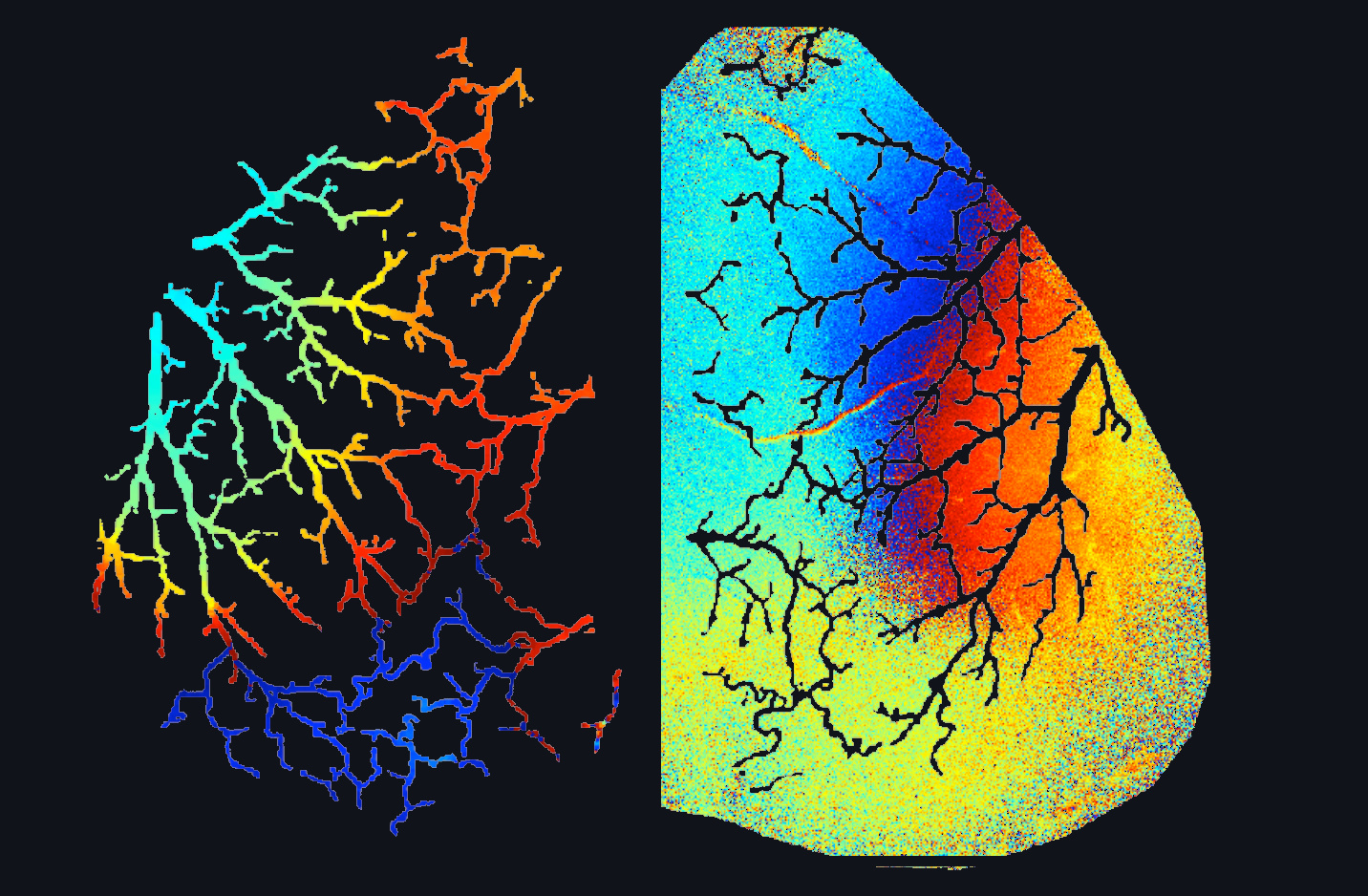
Vascular network of the mouse cortex colour-coded by vasomotion phase, showing long-wavelength traveling waves
Traveling waves of vasomotion. Penetrating arterioles of the cortex, colour-coded by oscillation phase — waves propagate at ≈2 mm/s and modulate perfusion by 20%.
0
Publications
0
Lead-authored
0
Imaging modalities
0
Continents, one lab
Scroll

Our mission

From the operating theatre to the bench — and back again.

Our laboratory is dedicated to advancing the understanding and treatment of neurosurgically relevant disease. As part of the Department of Neurosurgery at University Hospital Frankfurt, we work at the point where clinical observation and basic biology meet.

Disease exploration

Investigating the mechanisms behind brain metastasis, glioblastoma and neurovascular disorders — always anchored to a clinical question.

Advanced imaging

Confocal, two-photon and wide-field microscopy — including adaptive optics — to examine the brain at cellular and system scale.

High-dimensional analysis

Single-cell and high-dimensional expression profiling of patient-derived tissue, benchmarked against mouse models.

Translation to therapy

Working with clinical teams so discoveries reach practice — and so clinical observation keeps steering the laboratory.

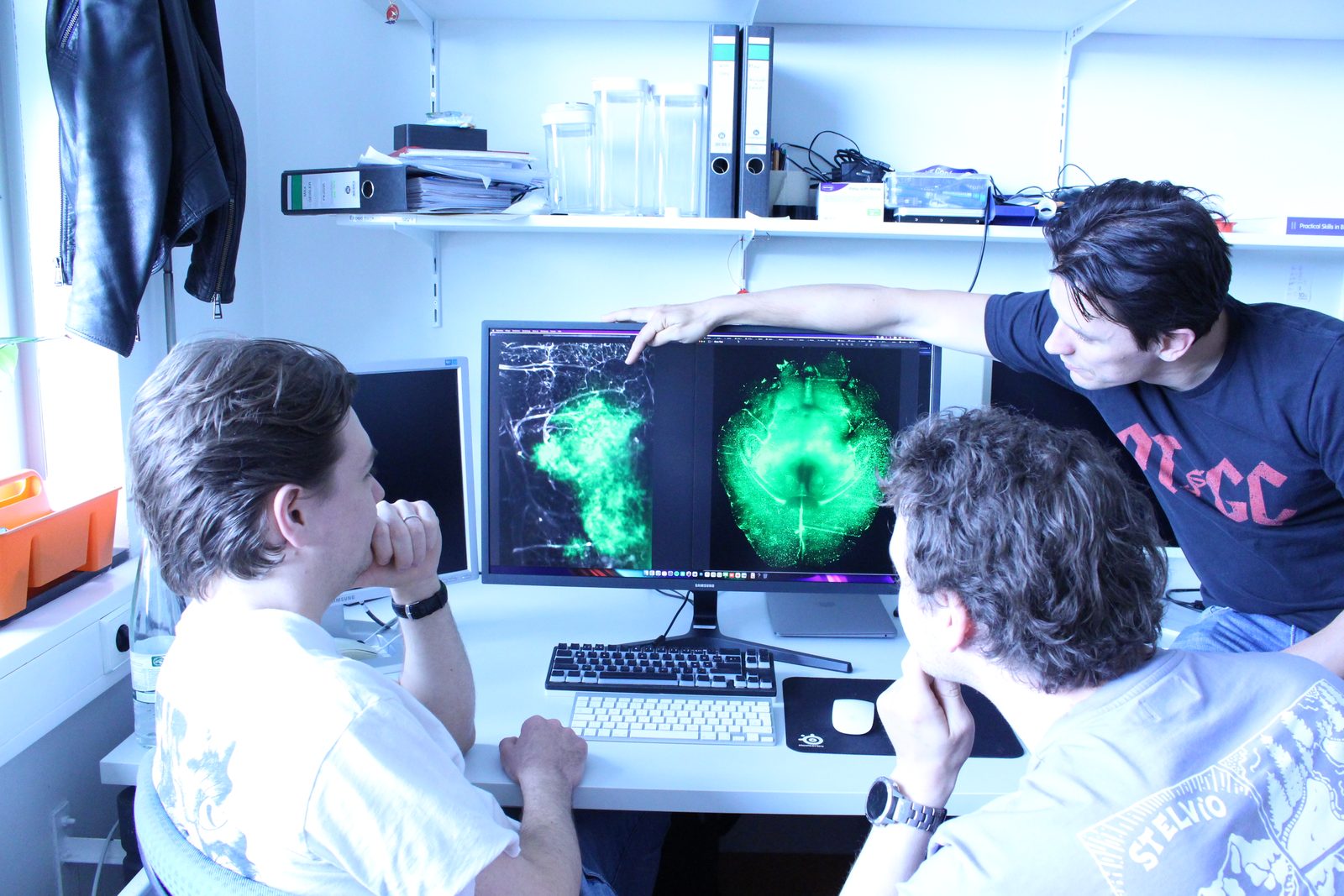
Members of the laboratory reviewing fluorescence imaging data of cerebral vasculature on screen

Research programme

Four questions we keep returning to.

Each of our lines of work sits at the interface between tumour biology and the cerebral vasculature — the tissue that decides whether a therapy ever reaches its target.

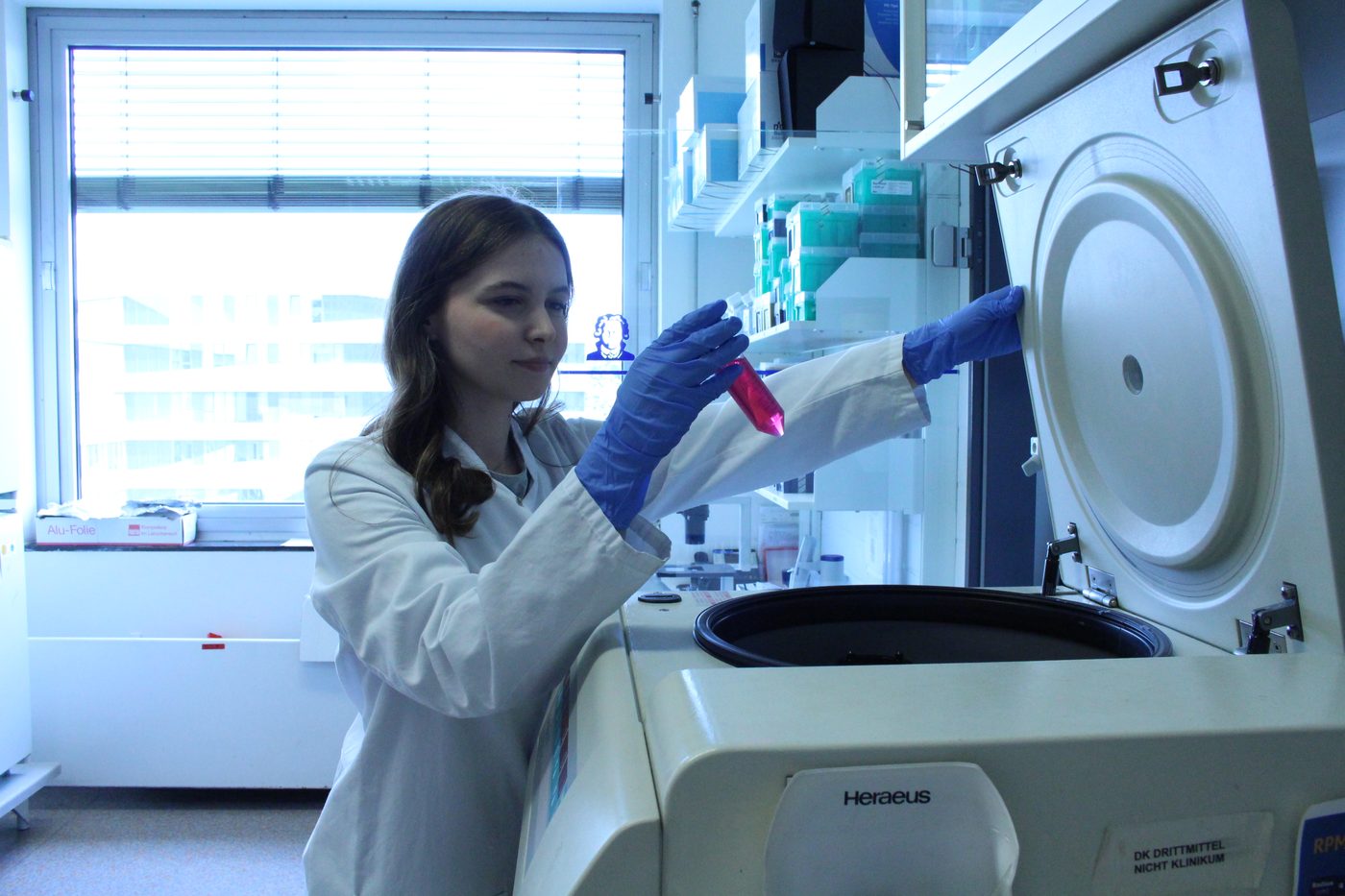
01 Preparation of patient-derived tissue samples in the laboratory

Brain metastasis & the pre-metastatic niche

Before a tumour cell ever arrives, the brain changes. We map metastatic and pre-metastatic tissue at single-cell resolution to find biomarkers that predict — and might prevent — brain and bone metastasis.
Single-cellPatient tissueBiomarkers
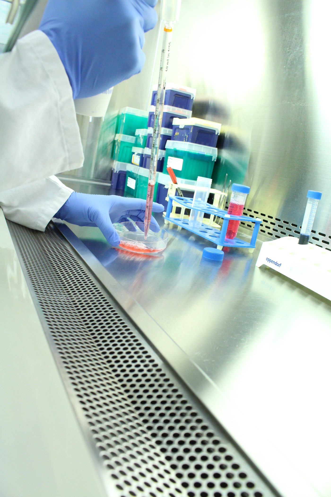
02 Pipetting under a laminar flow hood during a cell-culture experiment

Glioma biology & therapy resistance

Anti-angiogenic therapy works — until it does not. Our work on Ephrin-B2/EphB4 signalling and vascular architecture asks why gliomas escape treatment.

EphrinB2 · EphB4Anti-angiogenicsGlioblastoma
03 Phase map of cortical vasomotion showing traveling waves across the arteriolar network

Vasomotion & the neurovascular interface

Arterioles oscillate roughly once every ten seconds, independently of the heartbeat. We showed these oscillations form long-wavelength traveling waves that modulate cortical perfusion.

Cerebral blood flowTraveling wavesfMRI biology
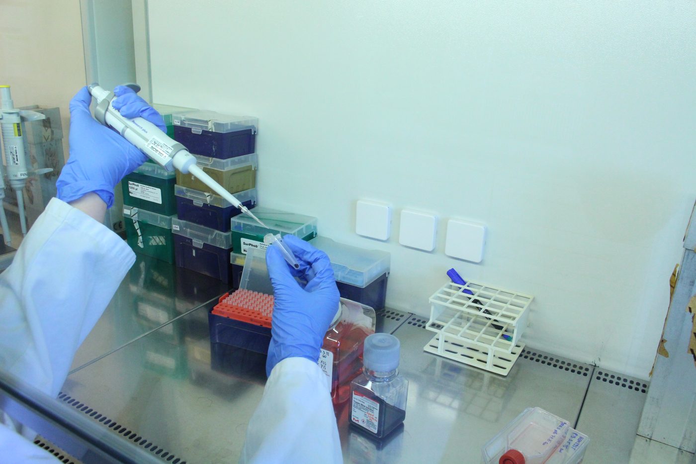
04 Multichannel pipette and reagent rack inside a laboratory hood

Optical methods & small-animal microsurgery

Better answers need better instruments. We build and apply adaptive-optics two-photon microscopy, chronic cranial windows and wide-field imaging.

Two-photonAdaptive opticsCranial windows
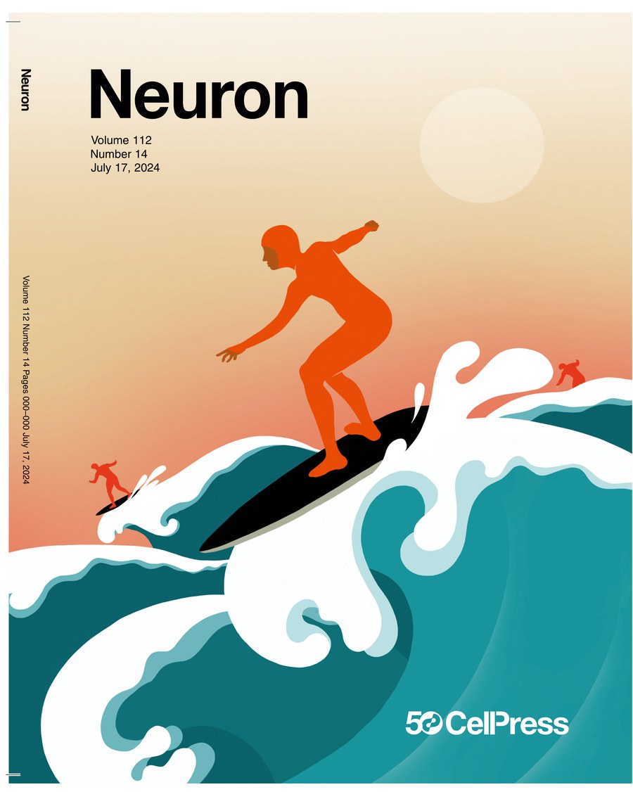
Cover of Neuron, Volume 112, Number 14, July 17 2024

Neuron 112(14) · 17 July 2024

Featured work

A washing machine for the brain.

Neuron 2024 Goethe University × UC San Diego

Using a combination of microscopic and biophysical methods, we visualised for the first time that the arterioles diving into the cerebral cortex pulsate independently of the heartbeat and of brain activity. These oscillations alter blood flow by 20% — and the effect is stronger at rest than in active brain regions.

The oscillations generate long-wavelength traveling waves that sweep along every arteriole at about two millimetres per second, in many different directions. The bulges they create on the vessel wall may help mix the cerebrospinal fluid bathing the brain, improving the clearance of misfolded proteins and metabolic waste — a mechanism that is disrupted in Alzheimer's disease and other neurodegenerative disorders.

“In further work here in Frankfurt, we will investigate how the traveling waves are altered during strokes, cerebral haemorrhages and neurodegenerative diseases, and what influence they have on the development of these diseases.” Thomas Broggini — first author, in the Goethe University press release, 31 July 2024

The team

Clinicians, biologists and physicists in one room.

Our interdisciplinary group brings together neurosurgical practice, molecular biology and optical physics — because none of these questions belongs to a single discipline.

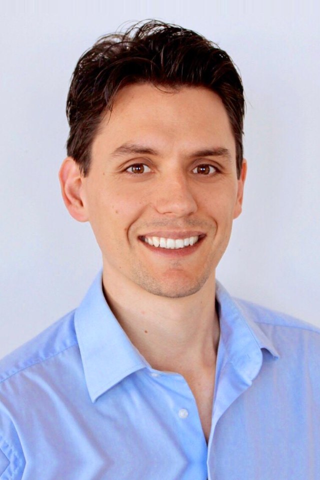
Portrait of Thomas Broggini

PD Dr. Thomas Broggini, PhD

Principal Investigator · Head of Laboratory · Working Group Leader

Thomas Broggini leads the Translational Neurosurgery Laboratory at the Department of Neurosurgery, University Hospital Frankfurt. His work combines optical imaging, small-animal microsurgery and advanced sequencing to understand how metastasis forms — in mouse and in patients — and how the cerebral vasculature governs what reaches the brain.

His route into science was not the usual one: he trained first as a chemistry laboratory technician in Switzerland, worked in industry, and only then studied biotechnology — an apprenticeship-first path he still credits for how he approaches an experiment.

Get in touch

Come and work with us.

Collaboration is at the heart of our mission. Whether you are a clinician, a researcher, or a student looking for a thesis project, we would like to hear from you — we host project weeks, practical seminars, MD and Master theses, and doctoral work.

Address
Neuroscience Center, House 89
Klinik für Neurochirurgie
Universitätsklinikum Frankfurt
Heinrich-Hoffmann-Str. 7
60528 Frankfurt am Main, Germany
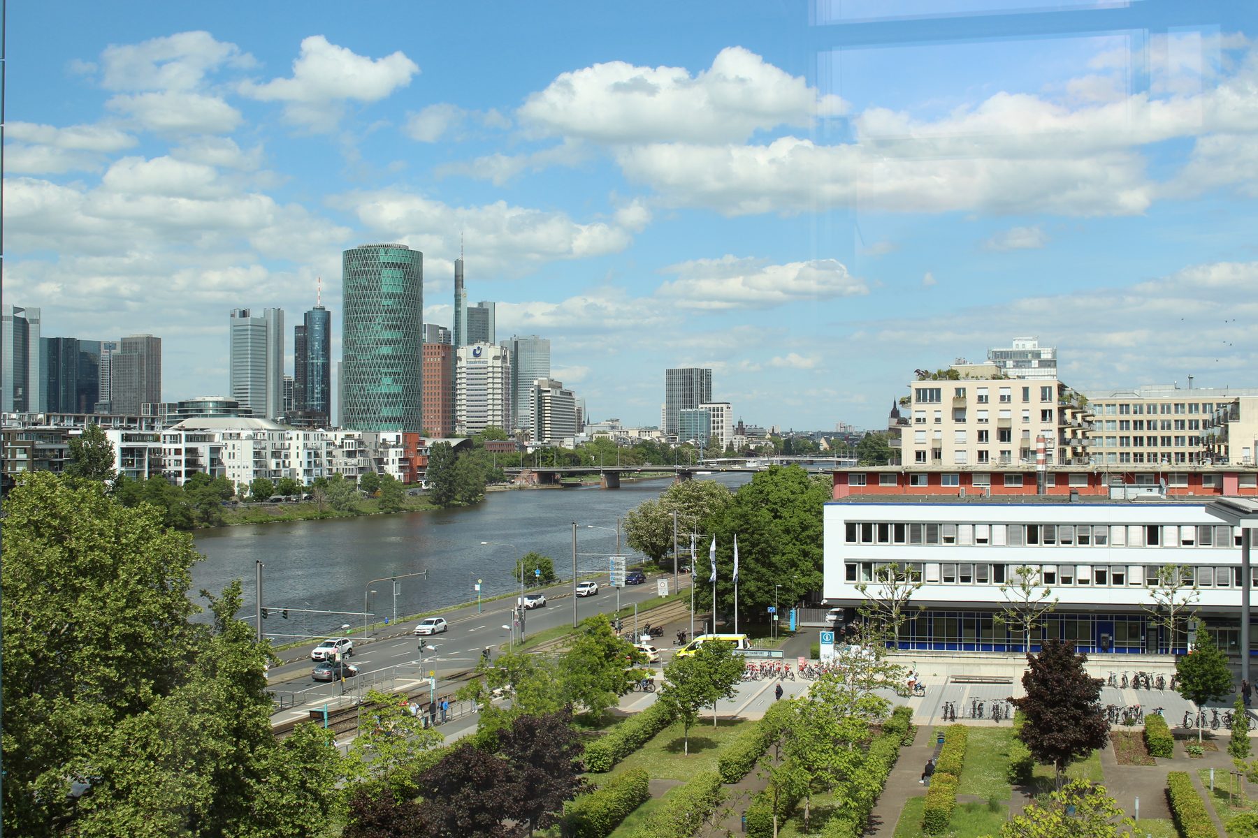
The University Hospital Frankfurt campus with the Main river and the Frankfurt skyline behind it

The campus on the Main — the skyline that gives our lab its mark.